Figure 13 shows that the N-1 peak was slightly better
resolved with the TSKgel SuperQ-5PW (20) than with the
TOYOPEARL GigaCap Q-650S, perhaps due to the smaller
particle size of the TSKgel resin. HPLC analysis of fractions
taken across the peaks (data not shown) revealed that
both resins were able to adequately resolve the full length
oligonucleotide.
PEGylated proteins
Ion exchange resins are frequently used for the purification
of pegylated proteins. Figure 15 shows the breakthrough
curves of five TOYOPEARL cation exchange resins for
mono-pegylated lysozyme.
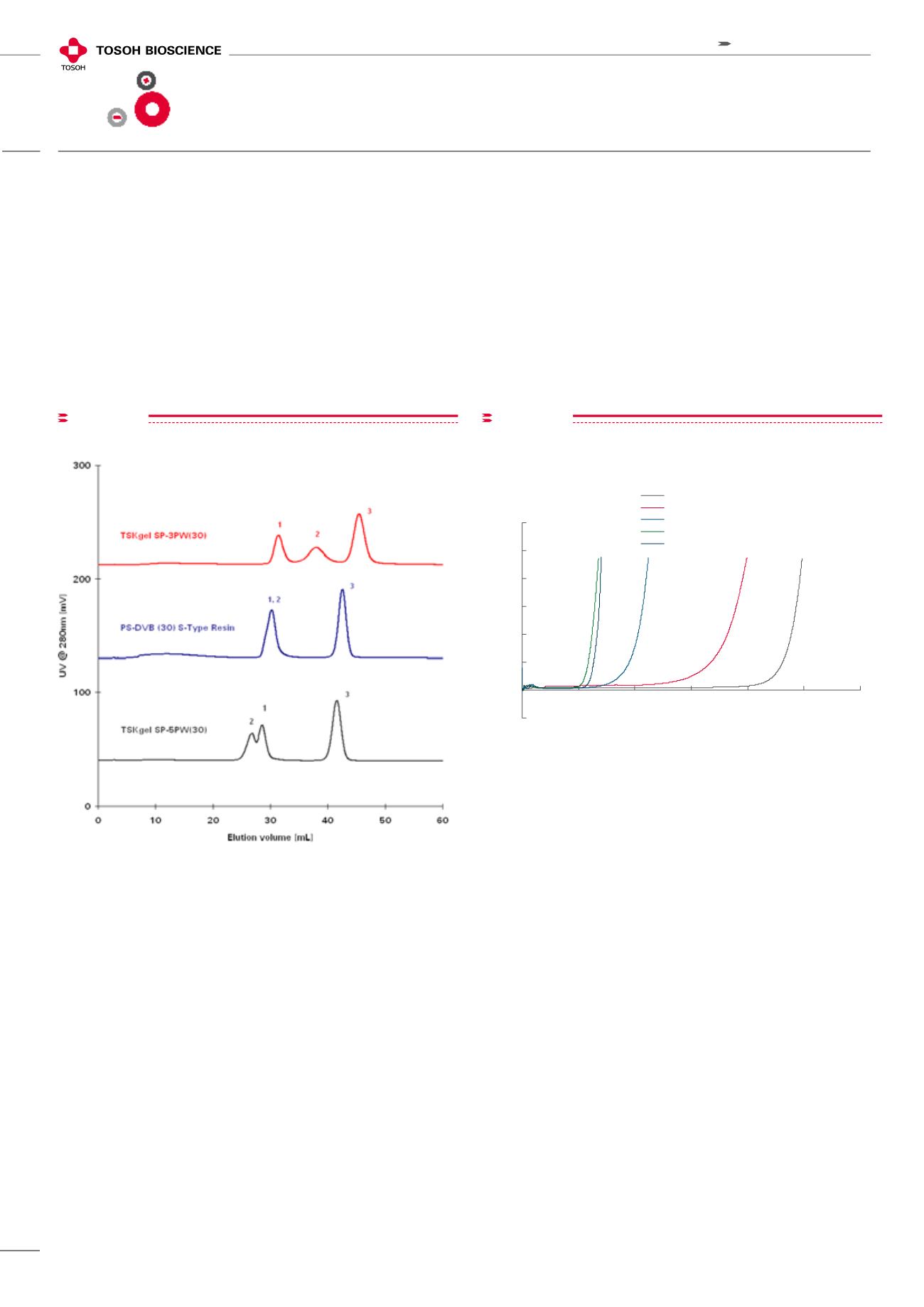
figure14
Column: 7.5 mm ID x 7.5 cm L; Mobile phase: A: 20 mM sodium citrate buffer
(pH 3.2)/ethanol = 8/2 (v/v); B: 1.0 mol/L NaCl in 20 mM sodium citrate buffer
(pH 3.2)/ethanol = 8/2 (v/v); Gradient: 60 min linear gradient from Buffer A
to Buffer B; Flow rate: 1.0 mL/min;
Detection: UV @ 280 nm; Temperature: RT;
Sample: 1. trypsinogen, 2. insulin, 3. lysozyme; 100 µL (0.5 mg/ml each)
Selectivity Comparison
figure15
-2
0
2
4
6
8
10
12
0
20
40
60
80
100
120
Mono-PEGylated Lysozyme Load (mg/mL-gel)
% Breakthrough
Toyopearl GigaCap S-650M
Toyopearl GigaCap CM-650M
Toyopearl SP-550C
Toyopearl SP-650M
Toyopearl CM-650M
Breakthrough curves of Mono-PEGylated Lysozyme
using Toyopearl cation exchange resins
Dynamic binding capacities were determined
at 10% breakthrough
Column size:
6mm ID x 40mm
Sample:
mono-PEGylated lysozyme
Loading buffer: 20mmol/L phosphate buffer (pH=7.0)
Elution buffer:
20mmol/L phosphate buffer (pH=7.0)
+ 0.5mol/L NaCl
Linear velocity: 212 cm/hr
Detection:
UV @ 280 nm
PEG MW= 5kDa
Dynamic binding capacities were determined at 10% breakthrough
Column size: 6 mm ID x 40 mm L; Sample: mono-PEGylated
lysozyme; Loading buffer: 20 mmol/L phosphate buffer (pH= 7.0)
Elution buffer: 20 mmol/L ph sphate buffer (pH= 7.0) + 0.5 mol/L NaCl
Linear velocity: 212 cm/h; Detection: UV @ 280 nm PEG MW= 5kDa
Breakthrough curves of Mono-PEGylated Lysozyme
using TOYOPEARL cation excha ge resins
28
IEC
Ion exchange
chromatography


